Animal MRI Facility
Mission of the Facility
The mission of the Biomedical Engineering Department in vivo high field Animal Magnetic Resonance Imaging (MRI) Facility is to provide  intellectual support and state of the art MRI facilities to researchers in the biomedical sciences and engineering who wish to image or collect nuclear magnetic resonance (NMR) spectra from small animals or biological samples with MRI.
This facility is not limited to in vivo imaging of small animals, but also supports MRI engineering development and other biomedical science projects that may require MRI or NMR spectroscopy.
Location and Contact Information
The facility is located 2360 Bonisteel Blvd, in room 1032 of the Bonisteel Interdisciplinary Research Building, which also houses the Functional MRI Laboratory.
Scientific and administrative questions regarding the facility should be directed to Dr. Luis Hernandez-Garcia.
Equipment
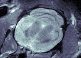
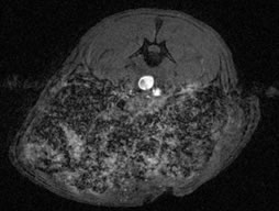
The Animal MRI Facility is equipped with a 7 Tesla MRI scanner from Agilent Technologies. The system was purchased using an NIH High End Instrumentation award (1 S10 RR 22974-01). This system has a 31 cm bore and has a 3 gradient system for larger animals, rats and mice, respectively, and supports a full range of MRI pulse sequences, including echo planar imaging and diffusion tensor imaging.
The system also includes 4 transmit channels for parallel excitation and 4 receive channels, along with a wide array of RF coils, including coil arrays for parallel imaging.
Full physiological monitoring, animal holders, rapid animal positioning systems, and an animal heater are available.
The control room to the scanner is also outfitted with a surgical table and sink for animal preparation as well as an exhaust system for anesthesia gasses. All the equipment necessary for small animal surgery, including an isoflurane vaporizer, a Harvard Apparatus rodent ventilator, a stereotaxic frame, a warming table, surgical instruments and physiological monitoring are available. 
The facility is operated by the users in collaboration with our faculty and students.
Use of the Facility
Use of the facility can be arranged for any investigator on campus with a legitimate research project.
To be granted access, investigators will need to:
- Succesfully complete a training course on the operation of the scanner. This course is offered twice a year and consists of two afternoon sessions.
- Sign up on the schedule posted at our C-Tools Site.
- In the near future, users will set up a recharge sub-account account in order to help defray the operation and maintenance costs of the facility. This will take the form of an hourly recharge rate, but this is not implemented. Please contact Luis Hernandez-Garcia to discuss this issue.
Managing Scan Data
Users will be responsible for transferring, archiving, and removing their data from the scanner when the experiment is complete, as disk space is limited and we do not provide a formal data archival system.
Collaboration Opportunities
We strongly encourage collaboration among our users. To facilitate this collaboration, we hold weekly meetings on Thursdays at 12:00 pm at the conference room, 1062 in the BIRB Building. These meetings are devoted to technical and administrative issues concerning operation of the scanner, but they are also intended to foster communication among researchers.
Facility C-Tools Site
To also encourage collaboration, we have developed a C-Tools Site is provided for users to exchange tips and protocols for use of the scanner. All users are encourage to contribute their knowledge to this site as they gain proficiency. Information on access to the C-Tools Site is provided to those inquiring about access to the facility.